You know your ABC’s, but what about your PCR? You know, Polymerase Chain Reaction. No, we’re not talking about nuclear meltdown. We are talking about DNA replication and amplification. This little machine carries out DNA replication and amplification.
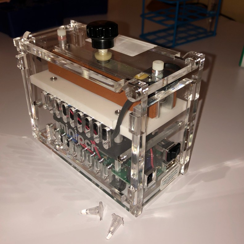
There are some necessary ingredients. For starts we need the DNA that we are going to amplify or “photocopy.” We call this the template. We need primers, which are little fragments of custom ordered DNA that will determine which part of the DNA template gets amplified. We need the enzyme (DNA polymerase) that creates the “photocopy.” The enzyme needs “toner” or “ink.” These are the little nucleotides: A, T, G, and C (the alphabet of our genetic code). The PCR thermal cycler will change temperature to make different steps of the reaction take place (usually 3 steps), over and over again. Each time it goes through the 3 steps, a cycle is completed. Take a calculator and enter 1 x 2, and hit “=”. You get 2. Hit “=” again, and you get 4. Again, 8. Each time you hit “=” it’s like completing a cycle, and the number doubles. Theoretically, if you started with one copy, and you hit the “=” button 40 times (or go through 40 cycles), you end up with 549,755,813,888 copies (about 550 billion copies) of the specific section of DNA we’re interested in. This all takes place in the teeny tiny tube in the photo. The amout of liquid you see in the tube is 20 microliters (1/3 of a drop of water). Of course, you can’t see the 550 billion copies of the DNA of interest with the naked eye.
Enter gel electrophoresis.
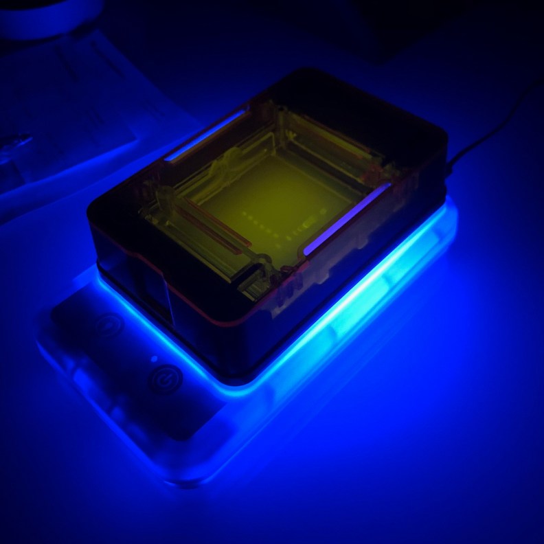
This machine holds a gel made of agarose (from seaweed). The agarose is like a dense forest of trees. Imagine the DNA as really long swimming pool noodles (20-100’s of feet long, or longer). Now imagine several individuals each holding a noodle and running through the forest as fast as possible. Those holding shorter noodles would get through the forest faster than those holding really long noodles. So we put the DNA (which has a very negative charge) in one end of the gel and apply an electrical current. The negative electrode is at the end of the gel closest to the DNA, and the positive electrode is at the opposite end of the gel. The DNA will get repulsed from the negative electrode (or be attracted towards the positive electrode) and travel through the gel. We can determine the length of the DNA by how fast (or slow) it migrates through the gel. The single bands in the video below represent photocopies of DNA that are 71 nucleotides long. In this case, that is what was expected, and the result suggests that the PCR reaction took place correctly and that the DNA template contained the area of interest (which was 71 nucleotides long). We can also cut out the DNA band and have it sequenced for definitive confirmation. If the template DNA did not have the area of interest, then nothing would have been amplified. The bands on the far right are DNA fragments of known lengths, and act as a “yardstick” to help us determine the length of the photocopied DNA (also known as, PCR product). The DNA glows because of a fluorescent dye in the gel.
PCR is the most precise way to detect mutations, contamination with certain bacteria, and genetically modified organisms.
